Journal of Structural Biology
Volume 120, Issue 1, 1997
ISSN 1047-8477
Supports open access
Based on images in article within by S M Spencer, J Y Sgro, K A Dryden, T S Baker, M L Nibert
(local PDF)
Click on images below to see larger versions.
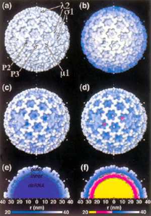
Cover based on Figure 2 d)
FIG. 2. The ISVP of mammalian reovirus type 1 Lang as determined by cryo-TEM and 3-D image reconstruction (Dryden et al., 1993). (a–d) Surface-shaded representations. The representations are identical except for the style of depth cueing that was imposed on the isosurface. Surface shading is applied to each representation as though a light were directed at the object from over the viewer’s left shoulder. (a) No additional depth cueing. Features representing outer capsid proteins l2, μ1, and s1 and channels P2 and P3 (Metcalf et al., 1991) are labeled. The μ1 protein is primarily present as fragments μ1N and μ1C in virions and as fragments μ1N, d, and f in ISVPs (Nibert and Fields, 1992). Only the base of the s1 fiber protein (Furlong et al., 1988) is visible in the virion and ISVP reconstructions (Dryden et al., 1993). (b) Z cueing. Color is a function of distance from the viewer. Hue remains constant, but brightness decreases as distance from the viewer increases. (c) R cueing, single-hue colormap. Color is a function of distance from the particle center (see color bar; r, radius, in nm). Hue remains constant, but brightness decreases as distance from the center decreases. (d) R cueing, stepped-hue colormap. Color is a function of distance from the particle center (see color bar; r, radius, in nm). A sharp transition in hue was introduced near radius 30 nm to approximate the boundary between outer capsid (blue) and inner capsid (magenta). Another sharp transition in hue was introduced near radius 23.5 nm to approximate the boundary between inner capsid (magenta) and genomic dsRNA(yellow). Across the radii of blue or magenta hue, brightness decreases as distance from the particle center decreases. (e, f) R-cued representations of the reovirus ISVP, cropped to remove the front half of the particle as viewed along a twofold axis. Only the top half of the cross section is shown. (e) Single-hue colormap, same as in (c). (f) Stepped-hue colormap, same as in (d).
Images by Stephan Spencer

