Book
The Coxsackie B Viruses
S. Tracy, N.M. Chapman and B.W.J. Mahy (Eds)
Springer 1997
ISSN 0070-217X
ISBN 3-540-62390-6
Library of Congress Catalog Card Number 15-12910
Click on images below to see larger versions.
Cover legends
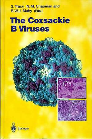
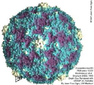
Coxscakievirus B3 image is a computer representation derived from X-ray coordinates (Mucklebauer et al. Structure 3:653, 1995). The PDB ENTRY for the virus is 1COV. The GRASP protein molecular surface is radially depth-cued to visually highlight surface features. The canyon is clearly evident. Note that the canyons are not continuous around the 5-fold icosahedral axes. (Courtesy of Dr. Jean-Yves Sgro, Institute for Molecular Virology, University of Wisconsin-Madison; sgro@rhino.bocklabs.wisc.edu jsgro@wisc.edu).
Photomicrographs of 0.6 micron thick hemotoxylin and eosin stained sections of heart (above) and pancreas (below) taken from a C3H/HeJ male mouse, 10 days post-inoculation with the cardiovirulent wild-type strain of CVB3, CVB3/DO. Note significant and widespread regions of necrosis and calcification in heart section typical of results induced by infection with a cardiovirulent CVB3. Pancreas has sustained severe damage to acinar cells. Original magnification x100.
Click on images below to see larger versions.

