Virology (Elsevier)
Volume 384, Issue 2, Pages 255-414 (20 February 2009)
Small Viruses, Big Discoveries: The Interwoven Story of the Small DNA Tumor Viruses
Edited by Paul F. Lambert (Archived version, Nov 23, 2020)
Click on images below to see larger versions.
Legend
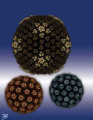
Journal Cover (Copyright © 2009): Shown are images of adenovirus (top center), polyomavirus (bottom right) and papillomavirus (bottom left). Images were created by Dr. Jean-Yves Sgro, Ph.D., Senior Scientist, Institute for Molecular Virology, University of Wisconsin-Madison with visualization software VMD. The following coordinate information was accessed through VIPERdb2. For adenovirus: PDB ID: 2bld; (Fabry et al., 2005, Embo J. 24:1645-1654); for polyomavirus: PBD ID: 1sid (Stehle and Harrison 1996, Structure 4:183-194); for papillomavirus: PBD ID: 1l0t (Modis et al., 2002, EMBO J. 21:4754-4762). For adenovirus, spike proteins are not visualized. For papillomavirus, the carbon-alpha backbone coordinates were converted to full coordinates with software Sybyl (Tripos, Inc.).

