VIROLOGY
Volume 445, Issues 1-2, Pages 1-244 (October 2013)
Special Issue: The Papillomavirus Episteme
(Archived Issue link, March 08, 2015)
Click on images below to see larger versions.
Legend
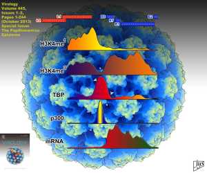
An HPV virion similar in structure to HPV18 (PDB 3IYJ - bovine papilloma) is shown radially colored to enhance the surface topography visual perception. Local plots derived from the reanalysis of ChIP-seq and RNA-seq datasets from HeLa-S3 cells show peaks in portrayed region of the HPV18 genome (red/blue gene map at top) for: Histone modifications, transcription factor binding, and RNA transcription. See Johannsen and Lambert for details.
Image Credits: UCSF Chimera virion image by Dr. J.Y. Sgro, UW-Madison | Reanalysis plots by Prof. Eric Johannsen
X-ray crystallography data: Wolf, M., Garcea, R.L., Grigorieff, N., Harrison, S.C. (2010) Subunit interactions in bovine papillomavirus. Proc. Natl. Acad. Sci. USA 107: 6298-6303

