Note: Scroll down for all maps
Archived version
Archived version of this page in Institute for Molecular Virology “virusworld” as it appeared on January 30, 2021.
History
These were the first “topographical” map representation of a viruse surface as if it were a “terrain” landscape on an exoplanet!
The topographical maps were created in the early 1990’s from Vsurf data on a Macintosh IIci with “Delta Graph 1.5” charting software that provided a better range of colors and 3D plotting capability compared to other software at the time.
However, the 3D angles of view were limited and to obtain this view the Vsurf data had to be “mirror imaged” and the resulting image was “mirror imaged” back.
Images were then edited to add text within Apple’s Claris MacDrawPro or Deneba’s Canvas 3.03.
BELOW is the original text minimally edited:
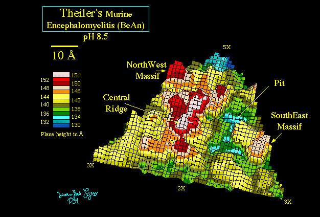
Topographical Maps
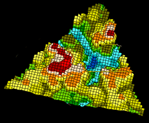
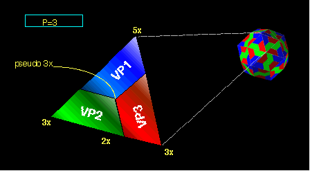
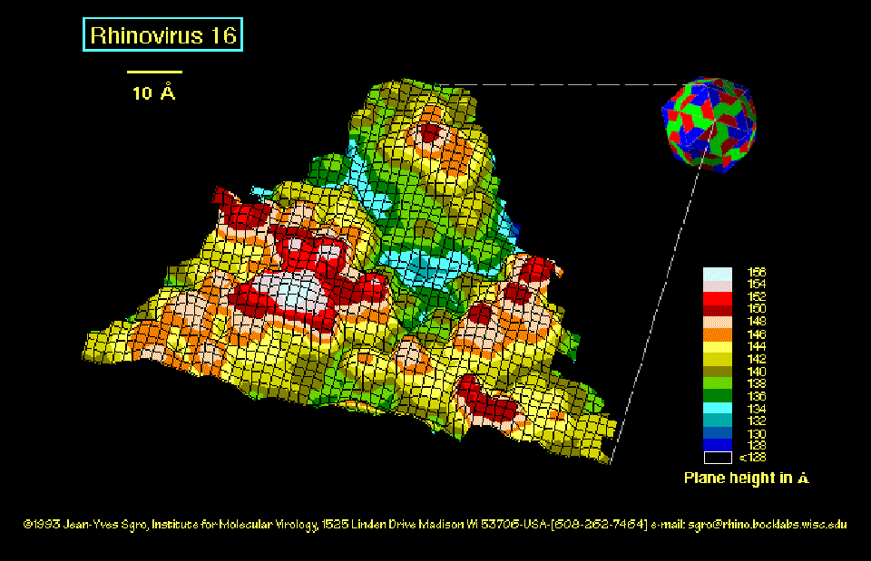
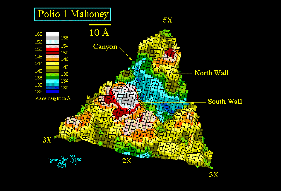
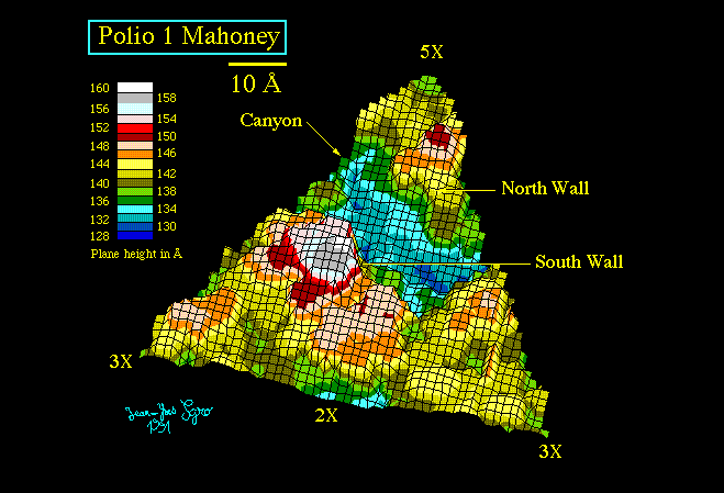
Topographical maps represent one “face” of the 60 faces of the icosahedron. The symmetryaxes would allow to reconstruct a complete icosahedral viral particle from one face.
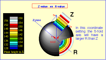
The topography is illustrated by coloring surface regions as a function of the distance to the center of the virus. That distance can be the radius (distance to the center) or a “Z” value (distance to a plane parallel to the icosahedral face).
The surface matrix is calculated with Vsurf (Rossmann and Palmenberg (1988) and rendered into Deltagraph on a Macintosh.
Topographical maps
File, general structure reference, PDB entry
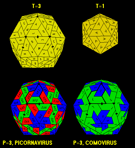
T=1,T=3,P=3 sketch
Canine Parvo Virus Tsao et al. (1991) 2DPV
Foot & Mouth Disease Virus (Q1 BFS 1860) Acharya et al. (1989) 1BBT
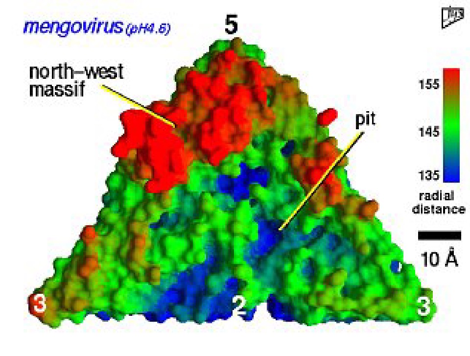
Mengo Virus (pH 4.6), Luo et al. (1987), 1MEC
Mengo Virus* (pH 4.6), Luo et al. (1987), 1MEC
Polio Virus Type 1 Mahoney Hogle et al. (1985), 2PLV
Polio Virus Type 3 Sabin Filman et al. (1989), -
Rhinovirus 14 Rossmann et al. (1985), 4RHV
Rhinovirus 16 Oliveira et al. (1993), -
Rhinovirus 1a Kim et al. (1990), 1R1A
Theilers Virus (BeAn Strain, pH 8.4) Luo et al. (1992), 1TMF
References
Program Vsurf
Rosssmann MG, Palmenberg AC (1988) Conversion of the putative receptor attachment site in picornaviruses. Virology 164, 373-382.
Additional reference:
Chapman MS (1993) Mapping the surface properties of macromolecules. Protein Science 2, 459-469
Virus structures
Acharya R., Fry E., Stuart D., Fox G, Rowlands D., Brown F. (1989) The three-dimensional structure of foot-and-mouth-disease virus at 2.9? resolution. Nature 37,709-716
Hogle J.M., Chow M, Filman DJ (1985) Three-dimensional structure of poliovirus at 2.9? resolution. Science 229,1358-1365
Kim S, Beoge U, Krishnaswamy S, Minor I, Smith TJ, Luo M, Scraba DG, Rossmann MG (1990) Conformational variability of a picornavirus capsid: pH-dependent structural changes of Mengo virus related to its host receptor attachment site and disassembly. Virology 175, 176-190.
Luo M, Vriend G, Kamer G, Minor I, Arnold E, Rossmann MG, Boege U, Scraba DG, Duke GM, Palmenberg AC (1987) The atomic structure of mengo virus at 3.0? resolution. Science 235, 182-191.
Luo M., He C., Toth K.S., Zhang C.X., Lipton H.L. (1992) The three-dimensional structure of Theiler’s murine encephalomyelitis virus (BeAn strain). Proc.Natl. Acad. Sci. USA 89, 2409-2413. (contains a topographical map: [Figure 2. color.] )
Filman DJ, Syed R, Chow M, Macadam AJ, Minor PD, Hogle JM (1989) Structural factors specificity in type 3 poliovirus. EMBO J 8, 1567-1579.
Oliveira MA, Lee W-M, Zhao R, Kremer M, Minor I, Rueckert RR, Diana GD, Pevear DC, Dutko FJ, McKinlay MA,, Rossmann MG (1993) The structure of human rhinovirus 16. Structure. 1,—,—. Submitted 1993.
Rotbart, H. A. and Kirkegaard K. (1992) Picornavirus pathogenesis: viral access, attachment and entry into susceptible cells. Semin. Virol. 3, 483-499. (contains a topographical map: [Figure 3 (A-F). color.] )
Rossmann MG, Arnold E, Erickson JW, Frankenberger EA, Griffith JP, Hecht H-J, Johnson JE, Kamer G, Luo M, Mosser AG, Rueckert RR, Sherry B, Vriend G (1985) Structure of human common cold virus and functional relationship to other picornaviruses. Nature 317, 145-153.
Tsao J., Chapman M.S., Agbandje M., Keller W., Smith K., Wu H., Luo M., Smith T. J., Rossmann M.G., Compans R.W. and Parrish C. (1991) The three-dimensional structure of canine parvovirus and its functional implications. Science 251, 1456-1464. (contains a topographical map: [Figure 1. color.] )
Source
Compiled July 1993 by:
Jean-Yves Sgro, Ph.D., Senior Scientist, Institute for Molecular Virology, Robert Bock Laboratories, University of Wisconsin-Madison, 1525 Linden drive, Madison Wi 53706 -USA.
Tel: 608-262-7464
Fax: 608-262-7414
e-Mail: jsgro@wisc.edu
Files are supplied “as is” and are screen captures of the original drawings which are polygon based images within Canvas 3.03 or MacDraw Pro (and are much bigger in byte size!). Files can be redistributed but not sold except for the price of medium and aknowledgment of source as well as the README file are provided.
(*) This images was created with program GRASP directly on Silicon Graphics. View is along the icosahedral 2-fold axis.


